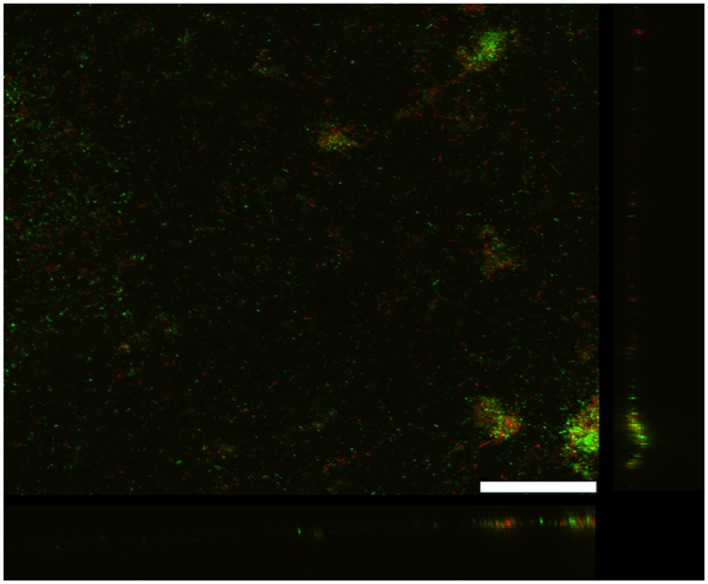
FIGURE 2.
Confocal scanning laser microscope image of an anode biofilm of G. uraniireducens that was producing 0.074 mA/cm2 current. Top-down three-dimensional, lateral side views (right image), and horizontal side views (bottom image) of cells stained with LIVE/DEAD BacLight viability stain. The size bar is 75 microns.

