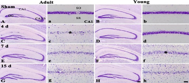
Figure 1.

CV staining in the hippocampus of the adult (left two columns) and young (right two columns) sham- (A, a, B and b) and ischemia-operated- (C – H and c – h) groups. In the adult ischemia-operated-groups, CV+ cells are damaegd in the stratum pyramidale (SP) from 4 days after ischemia/reperfusion (asterisk). However, in the young ischemia-operated-groups, CV+ cells are damaged from 7 days post-ischemia (asterisk). SO, stratum oriens; SR, stratum radiatum. Scale bar = 800 (A – H) and 50 (a –h) μm
