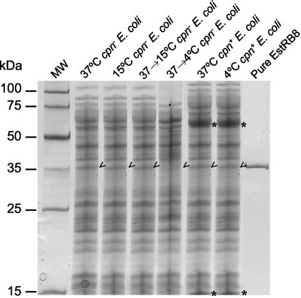
FIG. 1.
SDS-PAGE analysis of cell extracts of E. coli cpn and cpn+ strains. From left to right, the lanes contained molecular weight markers (MW), E. coli cpn cells grown at 37°C, E. coli cpn cells grown at 15°C, E. coli cpn cells induced at 37°C and then 15°C, E. coli cpn cells induced at 37°C and then 4°C, E. coli cpn cells grown at 37°C, E. coli cpn cells grown at 4°C, and pure esterase. The asterisks and arrowheads indicate the positions of Cpn60/Cpn10 and EstRB8, respectively. Each lane contained 10 μg of protein.

