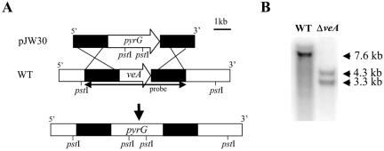
FIG. 1.
Deletion of the veA gene in A. parasiticus. (A) Diagram of the veA open reading frame and how it was replaced with the pyrG selectable marker from pJW30 by homologous recombination to generate the ΔveA mutant, TJW41.21. Solid bars indicate the 5′ and 3′ flanking regions of veA. (B) Southern analysis of wild-type (WT) strain and the ΔveA mutant strain. Genomic DNA was digested by PstI. A 6.1-kb PCR product containing veA was obtained with the primers 5′-AGA GAT GTC AAG TTC GAG TCG AG-3′ and 5′ CTC GGC ACC CAG CGT CAT CC 3′ and was used as a veA probe (indicated by arrows in panel A). Expected hybridization band patterns: wild-type strain, 7.6-kb band; ΔveA strain, 3.3- and 4.3-kb bands.

