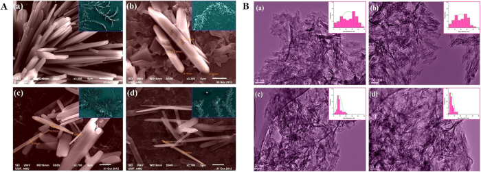
Figure 2.
(A) SEM images of ZnO nanoparticles prepared at different stirring conditions (a) 500 rpm, (b) 1000 rpm, (c) 1500 rpm and (d) 2000 rpm. The large colonies of the respective samples are shown in insets. (B) TEM images of ZnO nanoparticles prepared at different stirring conditions (a) 500 rpm, (b) 1000 rpm, (c) 1500 rpm and (d) 2000 rpm. Insets show the particle-size distribution curves.

