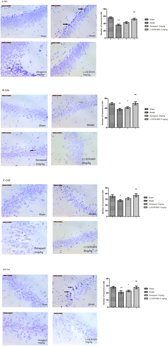
Figure 2. Typical neuropathological changes observed in the hippocampus 4 weeks after bilateral ligation of the common carotid arteries.
Neuronal loss, shrinkage (solid arrows) and vacuolation (hollow arrows) of neurons were observed in the CA1, CA3, CA4 and DG regions of the hippocampus in the model group rats. Long-term administration of (-)-SCR1693 and donepezil attenuated chronic hypoperfusion-induced neuronal damage to different degrees (scale bars = 50 μm). Histograms show the neuron number per field of view (×400) in the CA1, CA3, CA4 and DG regions (mean ± SEM, n = 6). *p < 0.05 vs. Sham group; #p < 0.05 vs. Model group. **p < 0.01 vs. Sham group; ##p < 0.01 vs. Model group.

