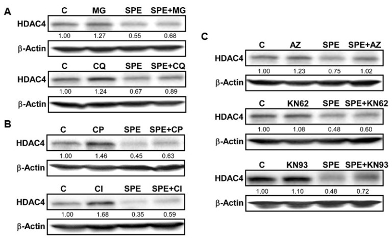
Figure 3.
Lysosomal and calpain-mediated degradation of HDAC4 by SPE. (A) Western blot analysis of HDAC4 in RAW 264.7 macrophages pretreated with 10 μg/mL of MG-132 (top), or 50 μM of chloroquine (bottom) for 1 h and then treated with 25 μg/mL of SPE in the presence of inhibitors; (B) Western blot analysis of HDAC4 in RAW 264.7 macrophages pretreated with calpeptin (10 μg/mL) for 1 h and then treated with 25 μg/mL of SPE in the presence of inhibitors; (C) Western blot analysis of HDAC4 in RAW 264.7 macrophages pretreated with CAMKII inhibitor KN-93 at the concentration of 5 μM for 1 h and then treated with 25 μg/mL of SPE in the presence of inhibitors. A represented blot of three independent experiments is shown.

