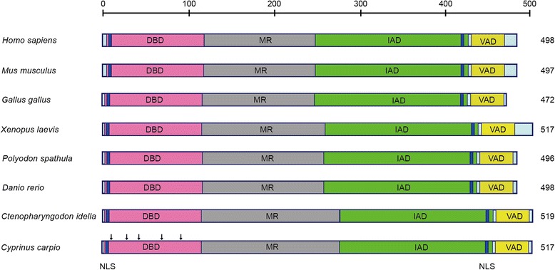
Fig. 1.

Domain organization of ccIRF5 and other vertebrate IRF5 proteins. The DBD (pink), MR (grey), IAD (green) and VAD (yellow) are depicted in different colours. The two NLSs are boxed in blue and the conserved tryptophan residues are indicated by downward arrowheads. Numbers refer to the length of the amino acid sequences. The accession numbers are listed in Table 2
