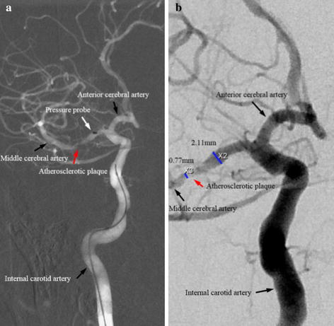
Fig. 1.

Digital subtraction angiographic images showing an atherosclerotic plaque in the middle cerebral artery (a image highlighting a microcatheter (marked by white arrow) and an atherosclerotic plaque in the middle cerebral artery (marked by red arrow); b image illustrating dimensions at the most stenotic site and the proximal disease-free site; a and b were from the same patient.)
