
Fig. 3.
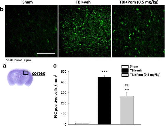
TBI induces neuron degeneration within the contusion region, and treatment with Pom significantly reduced the number of TBI-induced degenerating neurons. a Representative cresyl violet stained coronal brain section of the Sham (control without TBI) that shows the area of evaluation. b Representative photomicrographs showing the presence of FJC-staining at 24 h in the sham (control without TBI), TBI + veh group, and TBI + Pom administered 5 h post-injury group. c Quantitative comparison of mean densities of FJC-positive cells in the cortical contusion area at 24 h post-injury. There was a significant decrease in the number of FJC-positive cells in the TBI + Pom group. Mean ± S.E.M. (n = 5 in each group). **p < 0.01, ***p < 0.001 compared with the sham group. ## p < 0.01 compared with the TBI + veh group. Scale bar = 100 μm