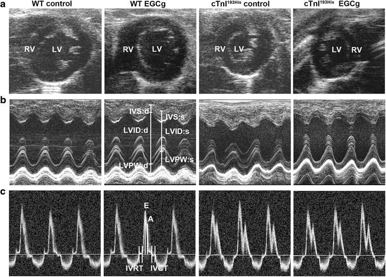
Fig. 1.

Determination of cardiac function with high resolution echocardiography in WT and RCM TG mice with or without treatment of EGCg. a Representative two-dimensional short axis views obtained from four different groups of the experimental mice. b Representative M-mode images and parameter calculation in experimental mice. c Representative images of pulsed Doppler of mitral inflow obtained from the experimental mice. LV left ventricle, RV right ventricle, E peak velocity of mitral blood inflow in early diastole, A peak velocity of mitral blood inflow in late diastole; E/A ratio; IVRT isovolumic relaxation time; IVCT isovolumetric contraction time, LVID:s left ventricular internal diameter end systole, LVID:d left ventricular internal diameter end diastole
