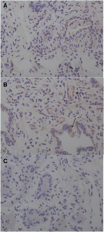
Fig. 3.

Sections of mammary gland immunostained for LAP (×400). a Alveolar epithelial cells from the LCD group that are immunonegative for LAP. b Alveolar epithelial cells from the HCD group that are immunopositive for LAP. Arrows show positive immunoreactions for LAP. c Alveoli cultured with normal rabbit IgG as controls for LAP
