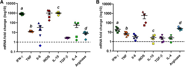
Fig. 3.

Gene expression of immunological markers in golden hamsters infected with Leishmania (V.) braziliensis. The graphics show the fold change of mRNA levels in skin (a) and lymph node (b) of golden hamsters infected with 105 Leishmania (V.) braziliensis promastigotes, 110 days post-infection. The relative quantification was performed by the comparative Ct method (△△Ct), using lymph node or skin from uninfected animals, respectively, as calibrator (Fold change = 1), as indicated by the dotted line. Each symbol represents a sample from a different animal, and each gene target is represented by a different colour. The median and interquartile intervals are indicated in the graphics. The letters a, b, c and d indicates statistical differences between mRNA levels in skin and lymph node (IFN-ɣ: Rank Sum Test: T (5,5) = 40, P = 0.008, TNF: t-test: t (8) = 3.72, P = 0.003, IL-10: Rank Sum Test: T (5,5) = 40, P = 0.008, arginase: t-test: t (8) = -3.69, P = 0.006)
