Figure 5.

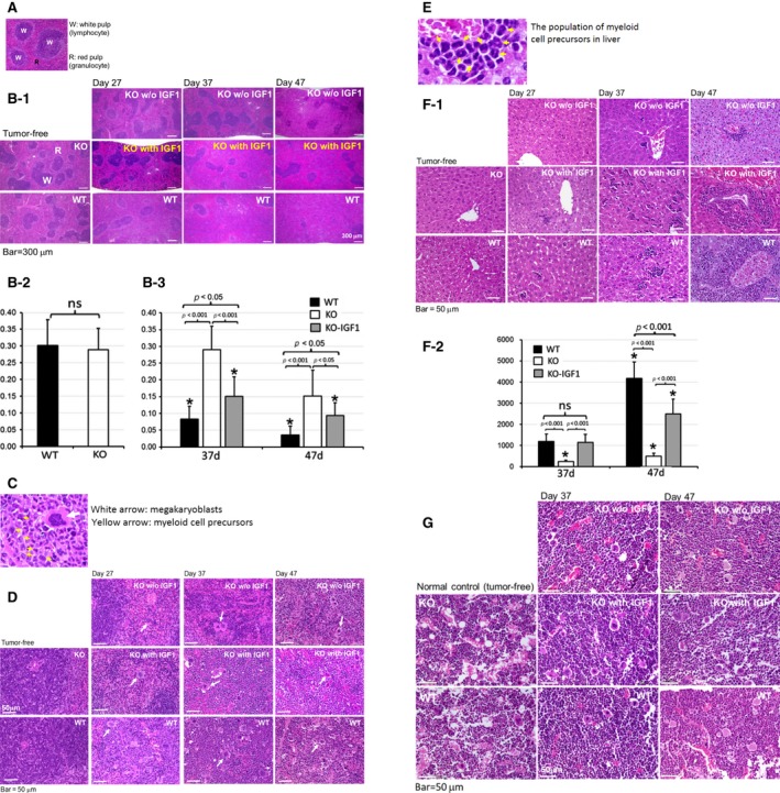
Microscopic images of spleen, liver, and bone marrow (BM) showed EphA4 deficiency reduced cancer‐related myeloproliferation. (A) microscopic image of white and red pulp of normal spleen. (B‐1) low magnification of the spleen with the expanded granulocyte‐rich red pulp and reduction of lymphocyte‐rich white pulp area on day 27, 37, and 47 of EphA4‐KO with or without IGF1 administration and control ‐WT tumor‐bearing mice. EphA4 deficiency reduced the splenic extramedullary hematopoiesis (EMH), which was enhanced by IGF1 treatment. (B‐2) histological quantification of the lymphocyte‐rich white pulp area percentage on tumor‐free mice (n = 4 each genotype). (B‐3) histological quantification of the lymphocyte‐rich white pulp area percentage on paired EphA4‐KO and ‐WT tumor‐bearing mice (n = 4* for each genotype and treatment, *the samples were collected on the indicated day or closely before or after the indicated day of Figure 5B‐3). (C) higher magnification showed the megakaryoblasts (white arrow) and myeloid cell precursors (yellow arrow). (D) EMH of megakaryoblasts and myeloid cell precursors of Figure 5B‐1 in higher magnification. (E) higher magnification showed myeloid cell precursors of liver. (F‐1) extramedullary hematopoiesis (EMH) was very rare in the liver of EphA4‐KO without IGF1 treatment (myeloid cell precursors were scarce in the liver of EphA4‐KO tumor‐bearing mice on day 27), but markedly enhanced when given IGF1 treatment on day 37 and 47 of tumor‐bearing mice. (F‐2) histological quantification of EMH cells, average number per high‐power field in the liver of paired EphA4‐KO and ‐WT tumor‐bearing mice. (n = 4* for each genotype and treatment, *the samples were collected on the indicated day or closely before or after the indicated day of Figure 5F‐2). (G) higher magnification of hematoxylin and eosin (HE)‐stained femur bone marrow (BM) section showed accumulation of myeloid cells at various stages of differentiation. Although cancer‐associated myeloproliferation of BM was identified on day 37 and 47 after tumor cell transplant, we could not be differentiated from EphA4‐KO tumor‐bearing mice with or without IGF1 treatment and control ‐WT tumor‐bearing mice.
