
Figure 3.
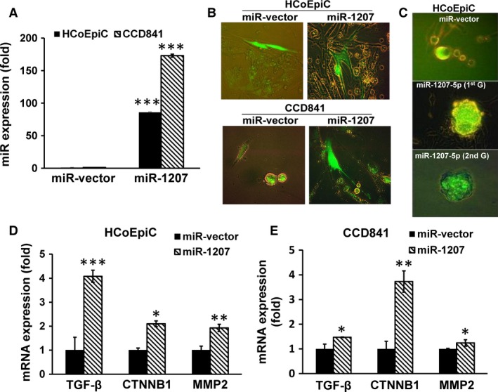
Forced expression of pre‐microRNA (miR)‐1207‐5p in HCoEpiC or CCD841 cells that caused a marked increase in miR‐1207‐5p levels (A), produced changes in morphology to epithelial mesenchymal transition phenotype as shown in the photomicrograph (B), formed spheroids following incubation for 14 days in medium containing serum [first generation; 1st G], which were disintegrated and the cells subsequently incubated in serum‐free stem‐cell medium for another 14 days [second generation; 2nd G] (C), accompanied by significant increases in transforming growth factor‐β‐receptor, Catenin (cadherin‐associated protein)‐β‐1 and MMP2 expression (D and E). Photomicrographs were taken under 400× magnification using Olympus fluorescence microscope with digital camera and DP2‐BSW software. Where applicable, data are presented as mean ± SD of three separate experiments. *P < 0.05, **P < 0.01 and ***P < 0.001.