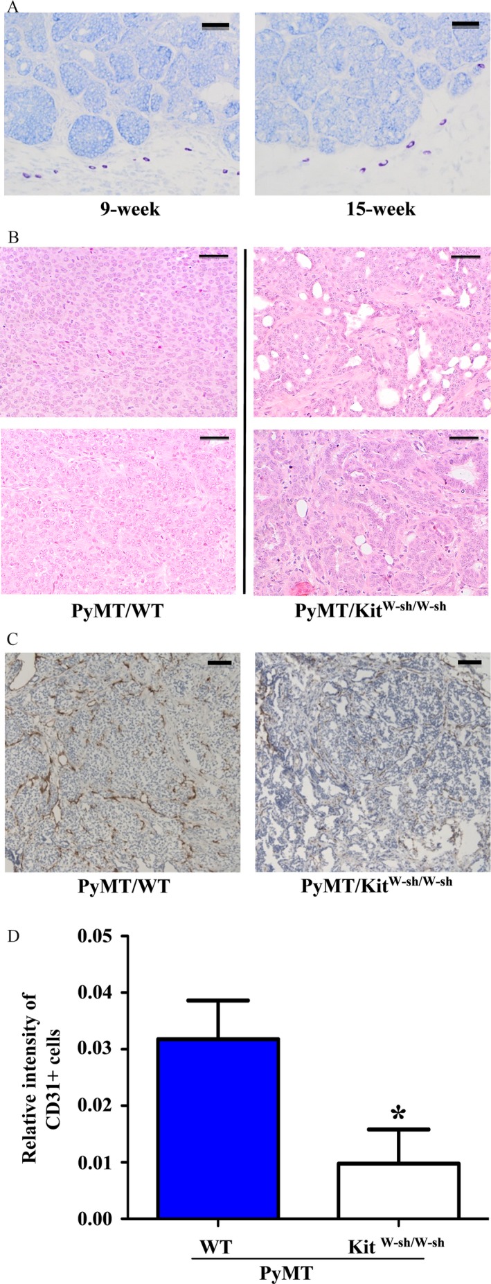
Figure 3.

Histological and immunohistochemistry analyses of mammary tumors. (A) Mast cells are present in the stroma of mammary tumors. Mammary tumors from 9 (left) and 15 (right) week‐old female PyMT/WT mice were dissected out, fixed, embedded, sectioned, and stained with Toluidine blue. Dark purple‐stained cells are mast cells. Bar = 100 μm. (B) Hematoxylin and eosin (H&E) staining of mammary tumors from PyMT/WT and PyMT/KitW‐sh/W‐sh female mice (10 weeks after tumor onset). Tumors from 2 PyMT/WT control mice displayed poorly differentiated morphology (left). In contrast, breast tumors from 2 PyMT/KitW‐sh/W‐sh mice showed more differentiated property (right). Bar = 100 μm. (C, D) Angiogenesis is impaired in tumors of PyMT/KitW‐sh/W‐sh mice. (C) Paraffin sections of tumors (2 months after tumor onset) from PyMT/WT and PyMT/KitW‐sh/W‐sh mice were stained with anti‐CD31 antibody. Bar = 200 μm. The dark brown colors are the CD31‐stained blood vessels. (D) The area stained by anti‐CD31 antibody was analyzed by the Image‐pro plus software as described in the Materials and Method section. Result shown is the average of relative CD31+ signal per microscope field from three tumors for each group. *P < 0.05.
