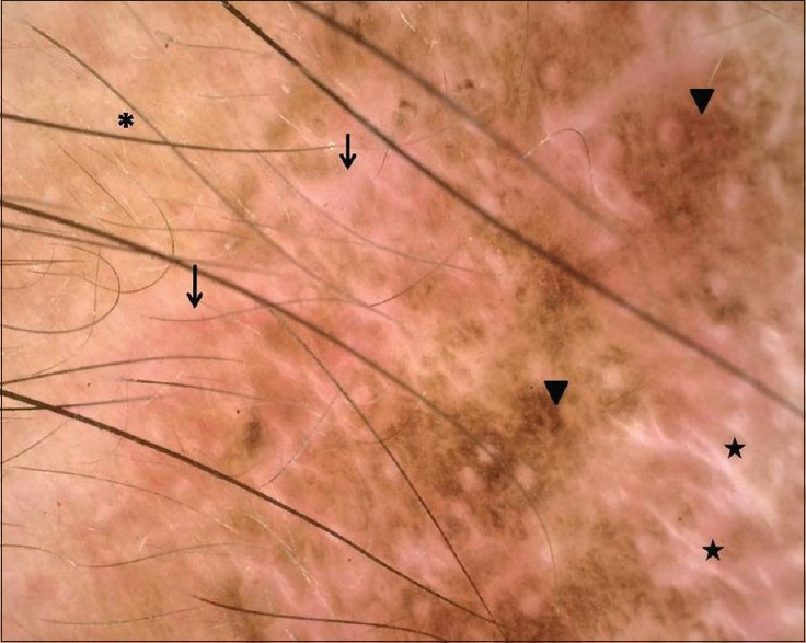
Figure 3.

Dermatoscopy of the residual lesion showing three zones – background erythema with dilated vessels (arrows) most prominent peripherally, followed by a prominent pigment network (arrowheads) and at the center chrysalis-like structures (stars), suggestive of new collagen induction at center. Note perilesional normal skin at upper left corner (Asterix “*”)
