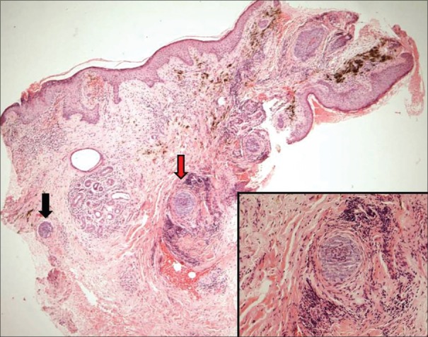
Figure 4.
Posttreatment biopsy-normal epidermis, lymphoid infiltrates in the dermis, and pigment incontinence in superficial dermis. Two tiny foci of basaloid cells in deep dermis (black and red arrows) surrounded by lymphocytic infiltrate and fibrosis representing immune zones (H and E, ×400). Inset: Close up of the foci (red arrow, ×1000)

