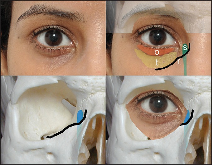
Figure 1.

The right eye demonstrating the tear trough anatomy (top left). Note the orbicularis roll (o) and the tear trough (t) in relation to the inferomedial orbital rim (r). The lacrimal sac (s) lies medially (bottom left and right), continuing below the orbital rim as the nasolacrimal duct. The ‘tear trough’ therefore does not lie over the orbital rim, but lateral to it
