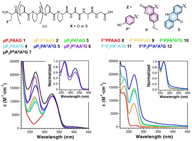
Fig. 1.
UV/Vis spectra of polyproline peptides containing multiple thioamides demonstrate non-additive changes in the thioamide absorption region (250–300 nm). All peptides contained multiple thioamide residues, varying numbers of prolines, and a 7-methoxycoumarin-4-ylalanine (μ) or p-cyanophenylalanine (F*) fluorophore. Left: Spectra of μ-containing peptides 1-7. The spectrum of a peptide containing two adjacent thioamides (5) is broadened compared to the spectrum of a peptide containing i, i+2 thioamides (6). Normalizing the absorbance at 270 nm makes clear the increased relative absorbance of 5 at longer wavelengths (Inset). Spectral broadening of the polythioamides can be even more clearly seen for peptides 8-12, containing F*. Particularly long wavelength features arising from thioamide-thioamide interactions are observed in the spectra of trithioamide peptides 7 and 12.

