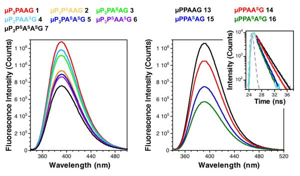
Fig. 2.
Increased fluorescence quenching by dithioamides observed in steady state spectra and fluorescence lifetime measurements. Left: Fluorescence emission spectra (λex = 333 nm) of peptides 1-7 at 5 μM in 10 mM sodium phosphate, 150 mM NaCl, pH7.0 buffer. Right: Fluorescence emission spectra (λex = 333 nm) of peptides 13-16 at 5 μM in 10 mM sodium phosphate, 150 mM NaCl, pH7.0 buffer. Inset: Time-correlated single photon counting measurements of (λex = 340 nm, λem = 393 nm) of peptides 13-16 at 5 μM in the same buffer, shown fit to single exponential decays (see ESI for details). Instrument response function is shown in light purple.

