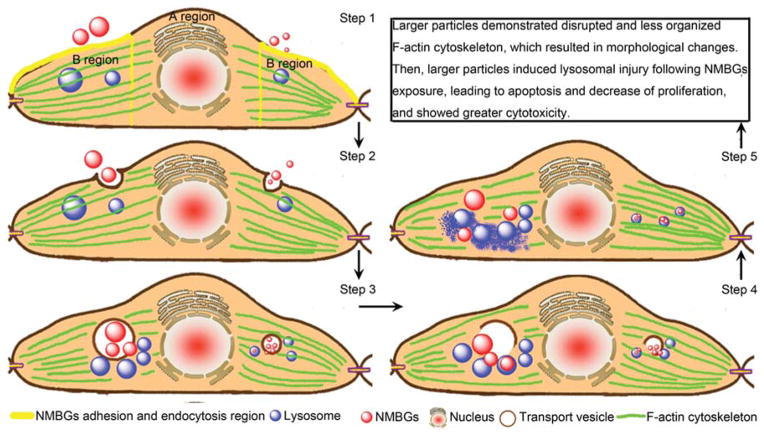
Figure 14.
Schematic illustration of the possible mechanism of NMBGs internalization in cells and the resultant cytotoxicity. The larger and smaller particles are shown on the left and right of the cell, respectively. In the first step, NMBGs are adsorbed on the perinuclear membrane of the cells. In the second step, transport vesicles serve as the major carrier of the internalized NMBGs for phagocytosis. In the third step, the internalized NMBGs carried by the transport vesicles induce the disruption and disorganization of F-actin cytoskeleton, which leads to the morphological changes. In the fourth step, the transport vesicles are broken and the particles come out of the broken vacuoles. Lastly, the smaller particles are encapsulated into the lysosomes and retained in the lysosomes; the larger particles may escape from the lysosomes and cause lysosomal damage, which will induce cell apoptosis.

