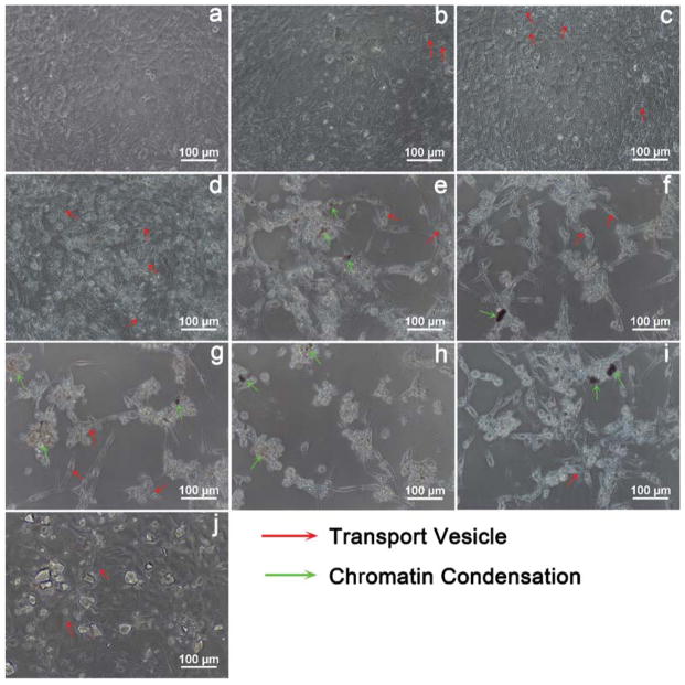
Figure 5.
The concentration of 100 μg/mL particles induced apoptotic morphology in MC3T3-E1 cells as revealed by light microscopy, (a) blank, (b) Group-61 nm, (c) Group-174 nm, (d) Group-327 nm, (e) Group-484 nm, (f) Group-647 nm, (g) Group-743 nm, (h) Group-990 nm, (i) Group-1085 nm and (j) Group-77 S. The groups shown in (e–i) induced apoptotic morphology with features such as filopodia formation and chromatin condensation.

