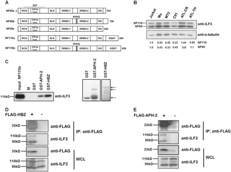
Fig. 1.

HTLV-1 HBZ and HTLV-2 APH-2 interact with NFARs in vitro and in vivo. (A) Schematic representation of NFAR isoforms. The functional domains are indicated and are as follows: RCN: region containing NES; EIF2α homology: region homologous to EIF2α; DZF: double zinc finger; NLS: nuclear localisation signal; DRBD-1/-2: double-stranded RNA binding domain −1 and −2; RG: arginine/glycine rich domain; GQSY: GQSY-rich region. (B) Expression levels of endogenous NF90 (90 kD) and NF110 (110 kD) in various cell lines. Equal amounts of whole cell extracts from Jurkat, HTLV-1 transformed cell lines, MT2 and C91, a HTLV-2 transformed cell line, Mo and ATL cell lines, ATL-TH and ATL-CR were electrophoresed on an 8% polyacrylamide gel and immunoblotted with antibodies directed against ILF3 which detects both NF90 and NF110, in addition to α-tubulin antibodies. For densitometry analysis of protein bands the individual signals for NF90 and NF110 in all cell lines were quantified and normalised against the signal for α-tubulin. To determine fold changes normalised values for HTLV and ATL cell lines were expressed relative to normalised values for uninfected Jurkat cells. The figures shown beneath the immunoblot denote the intensities of NF90 and NF110 relative to Jurkat cells (C) GST pulldown assays were performed by incubating purified NF110a with GST, GST-APH-2 or GST-HBZ immobilised on GST resin. The eluates were analysed by immunoblot with anti-ILF-3 antibodies (left panel) and coomassie blue staining (right panel). NF110a input corresponds to 20% of total NF110a loaded to each pulldown. M indicates protein molecular-weight marker. The arrows indicate purified GST, GST-APH-2 and GST-HBZ used in the pulldown assays. (D–E) HBZ and APH-2 interact with endogenous NF90 and NF110 in vivo. 293T cells seeded in 60 mm cell culture dishes were transiently transfected with 8 μg pFLAG-HBZ (D) or pFLAG-APH-2 (E) expression vectors as indicated. Immunoprecipitations were performed using an anti-FLAG M2 resin and precipitates were analysed by western blot using anti-FLAG and anti-ILF3 antibodies. IP: immunoprecipitation; WCL: whole cell lysate.
