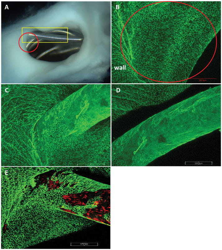
Figure 2. Aneurysm harvested at 8 weeks post embolization.
A, macro-photograph showing the coil loops at the neck orifice. Very thin membrane covering the coil loops at the peripheral area is observed (red circle). B, confocal microphotograph taken from the red circle area in A, showing the coil loops at the peripheral area in A within the red circle are completely covered with Cd31 positive cell ( large red circle area); those cells are confluent and continued up with the endothelial cells of the parent artery wall. C–D are taken from the rectangular area in A, showing the Cd31 positive cells covering on the coil surface not only limited to the periphery area,, extends to the center area. E, double staining for CD31 (green) and SMA (red), showing SMA positive cells are along with CD31 Positive cells on the coil surface at the peripheral area. (whole tissue mount staining: antibody for CD31 (A–D); double staining for CD31 and SMA (E), original magnificent 20X, water lens).

