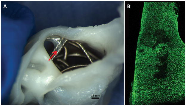
Figure 3. Aneurysm sample harvested at 8 weeks post embolization.
A, macro-photograph showing the membrane tissue between two coil segments at the center area (red arrow). B, confocal microphotograph showing the CD31 positive cells lining between the coil segments at the center area shown in A (red arrow) (whole tissue mount staining for antibody CD31, original magnification, 20x water lens).

