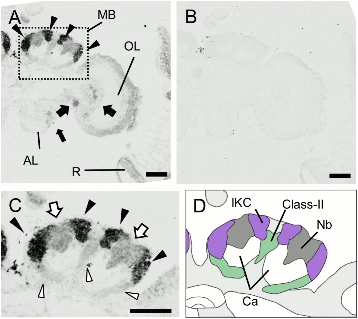
Fig 10. In situ hybridization of Mblk-1 in the pupal (P3) brain.
(A, B) Frontal sections of the right brain hemispheres hybridized with antisense (A) and sense (B) probes. Arrowheads and arrows indicate prominent signals in the MBs, OLs and ALs, respectively. MB, mushroom body; OL, optic lobe; AL, antennal lobe; R, retina. (C) Magnified view of the MB shown by a square in (A). Black and white arrowheads represent strong signals in the lKCs and weak signals in the class-II KCs, respectively. White arrows represent signals in the MB neuroblasts. (D) Schematic drawing of the brain structure seen in (C). The lKCs, class-II KCs, MB neuroblast clusters, and regions where somata of the other cells exist are colored by purple, green, grey, and light grey, respectively. Ca, calyces; Nb, MB neuroblasts; Ped, peduncles. Bars indicate 200μm.

