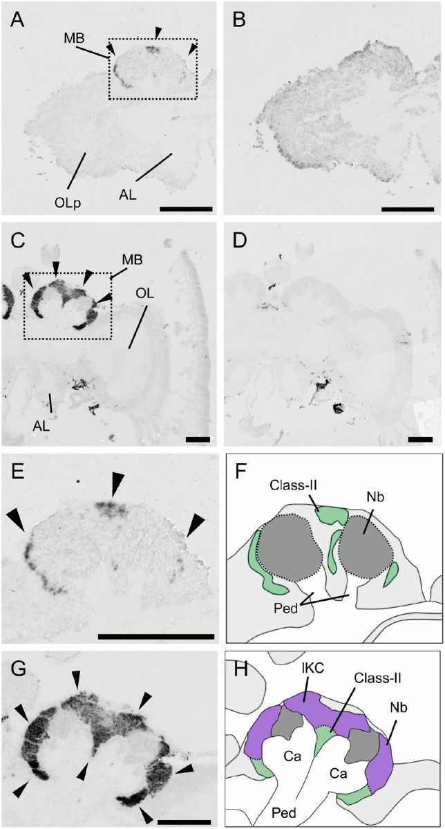
Fig 11. In situ hybridization of PLCe in the larval (L5F) and pupal (P3) brains.
(A-D) Frontal sections of brain hemispheres of hybridized with antisense (A and C) and sense (B and D) probes. (A and B) The larval (L5F) brain left hemisphere. (C and D) The pupal (P3) brain right hemisphere. Arrowheads represent prominent signals in the MBs. MB, mushroom body; OLp, optic lobe primordium; OL, optic lobe; AL, antennal lobe. (E-H) Magnified views of the MBs shown in (A) and (C) and their schematic views. (E and F) The larval MBs. (G and H) The pupal MBs. Arrowheads represent prominent signals in the MBs. The lKCs, class-II KCs, MB neuroblast clusters, and regions where somata of the other cells exist are colored by purple, green, grey, and light grey, respectively. Ca, calyces; Nb, MB neuroblasts; Ped, peduncles. Bars indicate 200μm.

