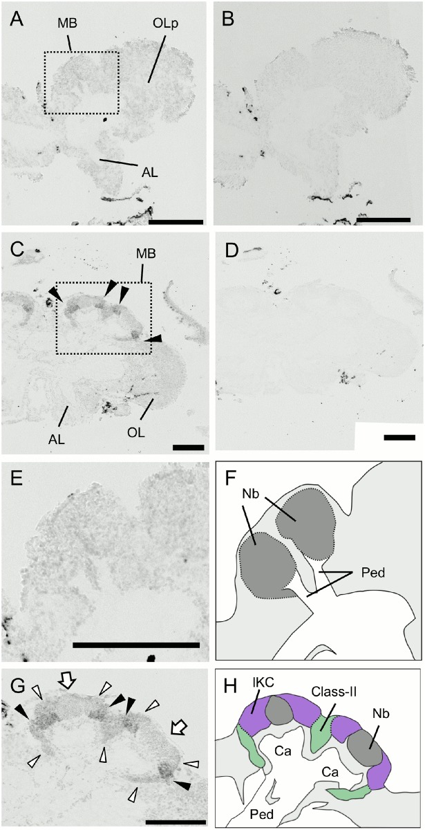
Fig 13. In situ hybridization of dlg5 in the larval (L5F) and pupal (P3) brains.
(A-D) Frontal sections of the brain right hemispheres hybridized with antisense (A and C) and sense (B and D) probes. (A, B) The larval (L5F) brain. (C, D) The pupal (P3) brain. Arrowheads in (C) indicate prominent signals in the MBs. MB, mushroom body; OLp, optic lobe primordium; OL, optic lobe; AL, antennal lobe. (E-H) Magnified views of the MBs shown in (A) and (C) and their schematic views. (E and F) The larval MBs. (G and H) The pupal MBs. Black and white arrowheads indicate moderate signals in the outer halves of the lKCs and weak signals in the remaining lKCs and class-II KCs, respectively. White arrows indicate weak signals in the MB neuroblast clusters. The lKCs, class-II KCs, MB neuroblast clusters, and regions where somata of the other cells exist are colored by purple, green, grey, and light grey, respectively. Ca, calyces; Nb, MB neuroblasts; Ped, peduncles. Bars indicate 200μm. The probes were prepared with the second set of primers mentioned in Materials and Methods.

