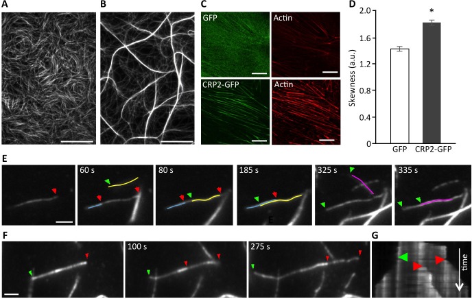
Figure 3. CRP2 promotes actin bundling in in vitro reconstituted assays and in breast cancer cells.
A. and B. Actin filaments (1 μM) polymerized alone (A) or in the presence of recombinant CRP2 (3 μM; B). C. Typical examples of ROI (13 × 13 μm) used for skewness measurements in Acti-stain 555 phalloidin-stained MDA-MB-231-luc cells expressing GFP alone or CRP2-GFP. D. Skewness average calculated from three independent experiments, including 200 optical sections as in (C). Error bars denote standard error. Significant level: *: p < 0.001. E. and F. Selection of images from Supplementary Movies 2 and 3 (real-time time TIRF microscopy) showing CRP2-induced crosslinking of actin filaments (E) and actin filaments elongation inside a bundle (E) and (F). In both cases 1 μM actin was copolymerized with 3 μM CRP2. Green and red arrows point to fast growing barbed ends of filaments elongating toward the left and right, respectively. For better readability some actin filaments were highlighted in color. G. Kymograph corresponding to (F) and Supplementary Movie 3, showing that CRP2 assembles bundles of mixed polarity. Bars = 20 μm (A and B), 10 μm C., 2 μm (E and F).

