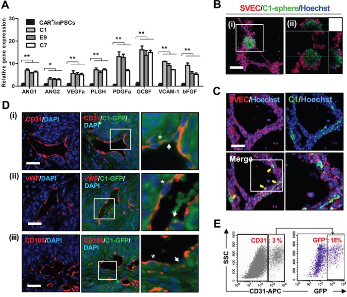
Figure 6. Angiogenesis potential of CAR+/mPSCsOct-4_hi.

A. Gene expression levels of proangiogenic factors in CAR+/mPSCs and CAR+/mPSCsOct-4_hi C1, E9, and C7 clones were analyzed using real-time PCR. Data are presented as the mean ± SD. * P < 0.05, ** P < 0.01, compared with CAR+/mPSCs. B. In vitro tube formation assay. SVEC4-10 cells and C1 clone-derived spheres, which stained with PKH26 and calcein-AM, respectively, were co-cultivated on Matrigel. The tube formation was recorded by fluorescence confocal microscopy for 8 h. C1 clone-derived spheres recruited SVEC4-10 cells to generate tube networks. C. A proportion of the C1 clone cells, indicated by arrows, were observed to integrate into the tube network. The magnified image of tube network depicts integration of the C1 clone cells. (Scale bar, 100 μm.) D. Immunofluorescence staining for the expression of endothelial antigens in C1-GFP clone-derived tumors. (i), CD31 was identified. (ii), vWF was identified. (iii) CD105 was identified. The magnified image shows that some GFP+ cells were involved in blood vessel formation (indicated by arrow), and a proportion of GFP+ cells also expressed CD31 (indicated by asterisk). (Scale bar, 100 μm.) E. CD31 expression in dissociated tumors of the C1-GFP clone was analyzed using flow cytometry. CD31+ sub-fraction, representing endothelial cells, constituted 3% of the whole tumor population. GFP expression was found in 18% of the CD31+ endothelial cell sub-fraction.
