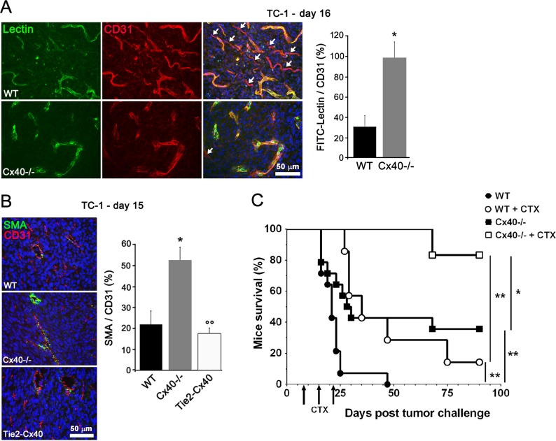
Figure 4. Cx40 deficiency causes enhanced tumor vessel maturation and functionality.
(A) Tomato lectin (green) labeled most of the vessels (identified in red by immunostaining for CD31) in tumors grown in Cx40−/− whereas some vessels were not stained by lectin in WT mice; white arrows indicate tumor vessels (CD31+) that were not perfused (lectin-). Data are mean + SEM of 8–10 fields from 3 different mice per group. (B) Immunostaining of alpha-SMA and CD31 showed the distribution of SMCs and ECs in the vessels of TC-1 tumors. The density of co-stained vessels was higher in the tumors induced in Cx40−/− than in those induced in Tie2-Cx40 and WT mice. Data are mean + SEM of 3–8 fields from 4–5 different mice per group. *p < 0.05 versus WT mice and °°p < 0.01 versus Cx40−/− mice. (C) Cyclophosphamide (CTX) treatment (arrows) of WT mice s.c. injected with TC-1 (n = 7, open circles) significantly prolonged mice survival (14% of surviving mice at the end of the 90 days experiment), as compared to WT untreated mice that all died within 7 weeks (n = 14; solid circles, p < 0.01). Survival of Cx40−/− mice injected with the same amount of TC-1 cells (n = 14; solid squares) was significantly higher than WT mice (36% of surviving mice, p < 0.01). Even more effective, CTX treatment in Cx40−/− mice (n = 6; open squares) resulted in 83% of the mice surviving (p < 0.01 as compared to CTX-treated WT mice). Significant differences by a log rank test are shown *p < 0.05 or **p < 0.01.

