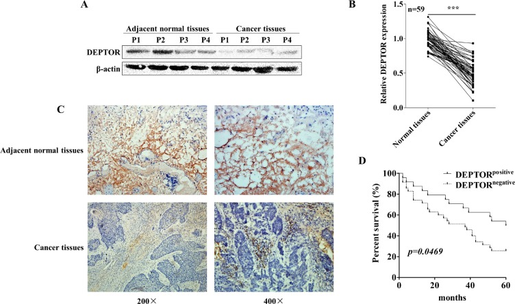
Figure 1. DEPTOR expression is decreased in human ESCC tissues and predicts a poor prognosis of ESCC patients.
(A) DEPTOR expression in ESCC tissues and non-cancerous adjacent tissues derived from human patients was detected by Western blotting, β-actin was used as the internal control. P1 means patient number 1. Western blotting results were quantified by ImageJ software and summarized in right panel. **p < 0.01. (B) mRNA expression of DEPTOR in samples from 59 ESCC patients was determined by qRT-PCR, GAPDH was used as an internal control and the date was analyzed by the 2−ΔΔCt method. ***p < 0.0001. (C) Representative pathological images showed expression of DEPTOR reduced in ESCC samples. (D) DEPTOR staining in all samples were photographed and scored and grouped into two groups by qualified pathologists in a blind manner. The Kaplan-Meier survival curve showed that DEPTOR positive group displayed a better prognosis that DEPTOR negative group.

