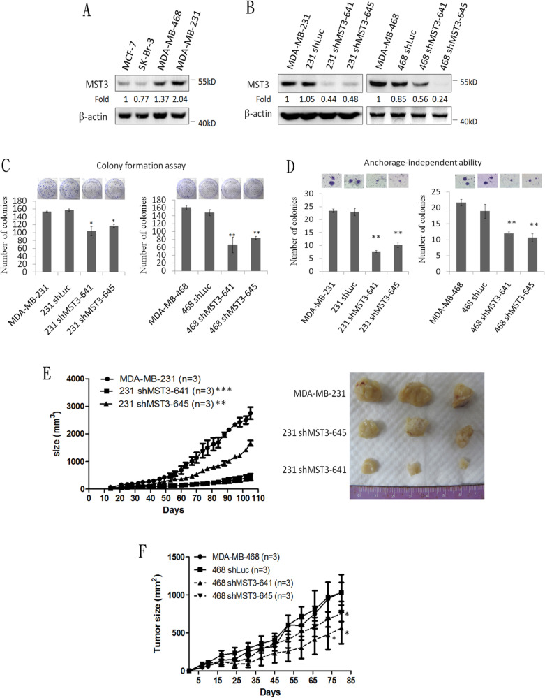
Figure 2. Attenuation of MST3 by shRNA inhibits proliferation, anchorage-independent growth, and tumor growth of breast cancer cells.
A. Western blotting analysis of MST3 in breast cancer cell lines. Equal amounts (30μg) of protein from whole-cell lysates were analyzed for MST3 and β-actin expression by Western blotting analysis. B. Western blotting analysis of MST3 in MDA-MB-231 and MDA-MB-468 cells that were stably transfected with two different MST3 shRNA plasmids (641 and 645). Equal amounts (30μg) of protein from whole-tissue lysates were analyzed for MST3 and β-actin expression by Western blotting analysis. C. The proliferation rates of the indicated cell lines were determined by colony formation assay. Representative images of colony formation assay were shown (upper panel). D. The ability of anchorage-independent growth of the indicated cell lines was determined by soft agar assay. Representative images of clone formation in soft agar were shown (upper panel). E. MST3 shRNA transfectants and MDA-MB-231 cells were injected s.c. into the flanks of NOD/SCID mice. After transplantation, tumor size was measured at the indicated days. Representative images of dissected tumors were shown (right panel). F. MST3 shRNA transfectants and MDA-MB-468 cells were injected s.c. into the flanks of NOD/SCID mice. After transplantation, tumor size was measured at the indicated days. Data are represented as mean ± SD from three independent experiments. * p < 0.05; **p < 0.01; *** p < 0.001.

