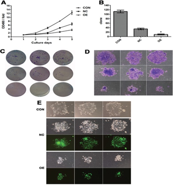
Figure 3. Increased expression of AB209630 suppressed the proliferation of FaDu cells.

A. The proliferation assay was performed on parental (CON), lentiviral vector control (NC), and overexpression lncRNA-AB209630 (OE) FaDU cells. The absorbance was measured on days 1, 2, 3, 4, and 5 according to the MTT method. B. The Gimsa-stained colonies were observed and measured under a microscope (×200). A bar graph shows the differences in colony formation among the three groups. The data are presented as the mean ± SD for three independent experiments (*P<0.01, compared with NC). C. Colonies were photographed. D. The diameter of OE sarcospheres was much smaller than that of CON and NC sarcospheres. E. Sarcospheres were photographed under a fluorescent microscope (×200).
