
Figure 1. Obatoclax reduced cell viability of both cisplatin-sensitive and –resistant esophageal cancer cells.
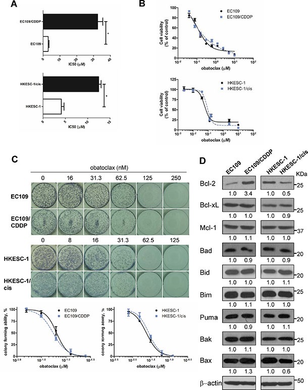
(A) Parental and cisplatin-resistant esophageal cancer cells (EC109 and its resistant subline EC109/CDDP, HKESC-1 and its resistant subline HKESC-1/cis) were treated with cisplatin (0–160 μM) for 48 h. Cell viability was then determined by MTT assay. IC50 values were calculated with Prism software. Data are presented as the mean ± S.E.M. from three independent experiments. *P < 0.05 compared with the respective parental cell line. (B) Cells were exposed to increasing concentrations of obatoclax for 48 h. Cell viability was then determined by MTT assay. Data are presented as the mean ± S.E.M. (n = 3) of a representative experiment performed in triplicate. (C) Cells were treated with obatoclax at the indicated concentrations for 48 h. Viable, adherent cells were counted and re-seeded (3,000 cells per well) into a well of a six-well plate (in triplicate), in the absence of obatoclax. Ten to twelve days later, colonies were fixed and stained. Each well shown is a representative image of at least nine similar wells (three independent experiments). Data are presented as the mean ± S.E.M. (n = 3) of a representative experiment performed in triplicate. (D) The basal expression levels of Bcl-2 family members were measured by Western blots. β-actin was used to evaluate protein loading. Blots were representative of 3 independent experiments. Quantification of the ratios of Bcl-2/β-actin, Bcl-xL/β-actin, Mcl-1/β-actin, Bad/β-actin, Bid/β-actin, Bim/β-actin, Puma/β-actin, Bak/β-actin, and Bax/β-actin is shown below each gel lane. The ratios were normalized to parental cell lines EC109 and HKESC-1 cells, respectively.