
Figure 2. Effects of obatoclax on apoptosis.
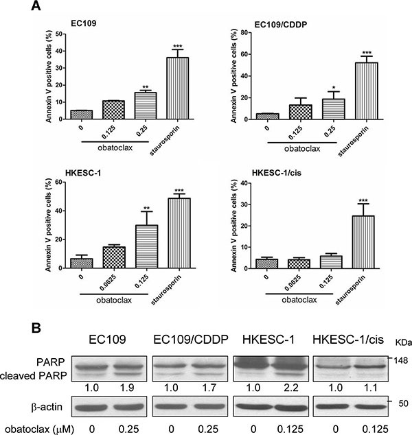
(A) Parental and cisplatin-resistant esophageal cancer cells (EC109 and its resistant subline EC109/CDDP, HKESC-1 and its resistant subline HKESC-1/cis) were exposed to the indicated concentrations of obatoclax for 48 h. Cellular apoptosis was assessed by phosphatidylserine (PS) externalization and binding of Annexin V-FITC. Data are presented as mean ± S.E.M. from three independent experiments. *P < 0.05, **P < 0.01, ***P < 0.001 as compared to the control. Staurosporin (50 nM) was used as a positive control. (B) Cells were treated with the indicated concentrations of obatoclax for 48 h. PARP cleavage was examined by Western blots. β-actin was used to evaluate protein loading. Blots were representative of 3 independent experiments. Quantification of the ratios of cleaved PARP/β-actin is shown below each gel lane. The ratios were normalized to control cells.