
Figure 7. Effects of obatoclax on lysosomal functions.
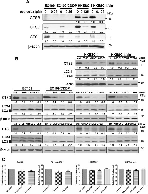
(A) Cells were treated with obatoclax at the indicated concentrations for 48 h. The active forms of cathepsin B, D and L were determined by Western blot analysis. Quantification of the ratios of CTSB/β-actin, CTSD/β-actin, and CTSL/β-actin is shown below each gel lane. The ratios were normalized to control cells. (B) The efficacy of cathepsin B (CTSB), cathepsin D (CTSD), or cathepsin L (CTSL) by respective siRNA was confirmed by Western blot analysis at 48 h post-transfection. Nontargeting siRNA was used as control siRNA, which has no homology to any known mammalian genes. Targeting CTSB, D, or L by RNA interference increased the conversion of LC3-I to LC3-II at 48 h post-transfection. Quantification of the ratios of CTSB/β-actin, CTSD/β-actin, CTSL/β-actin and LC3-II/β-actin is shown below each gel lane. The ratios were normalized to control siRNA-transfected cells. (C) Cell viability was determined at 48 h post-transfection of distinct siRNA targeting CTSB, D or L by MTT assay. The siRNAs used were CTSB1 siRNA, CTSD2 siRNA, and CTSL2 siRNA. Results were averaged and blots were representative of 3 independent experiments. *P < 0.05, **P < 0.01, ***P < 0.001 as compared to control siRNA transfected-cells.