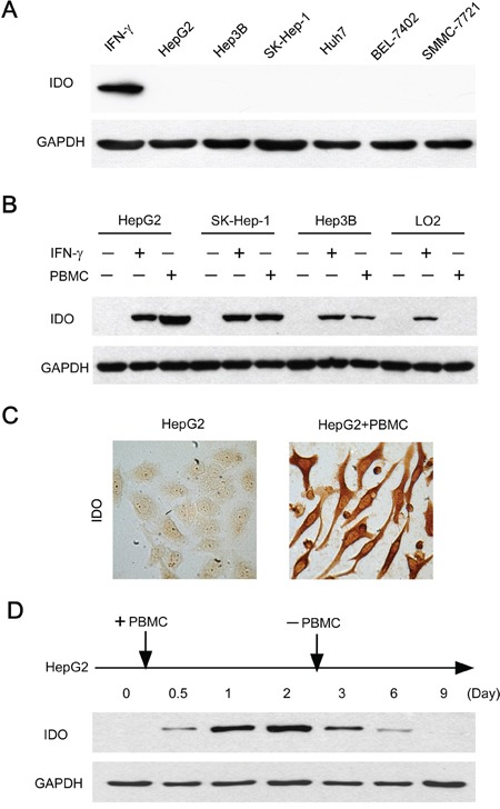
Figure 2. Immune cells contributed to the induction and maintenance of IDO1 in human hepatoma cells.

A. Human hepatoma (HepG2, SK-Hep-1, Hep3B, Huh7, SMMC-7721 and BEL-7402) cell lines did not constitutively express IDO1 in vitro. The IFN-γ treated HepG2 served as a positive control. B. Tumor cells (HepG2, SK-Hep-1, Hep3B) or normal liver cells (L02) were cultured with PBMC at a ratio of 1: 3 or IFN-γ (100 IU/ml) as IDO1-positive control for 2 days. For the coculture groups, PBMC were wash away before protein extraction. The expression of IDO1 protein in the attached cells was detected by western blot. C. HepG2 were cultured alone or with PBMC for 2 days. IDO1 protein was detected by immunocytochemistry. One of five representative areas is shown. D. PBMC were added to HepG2 at day 0, and were washed away after 2-day coculture. The IDO1 protein in the attached cells was detected by western blot at indicated times.
