
Figure 5. BRACO-19 specifically delocalizes TRF2 and POT1 from telomeres and induces telomeric 3′-overhang degradation.
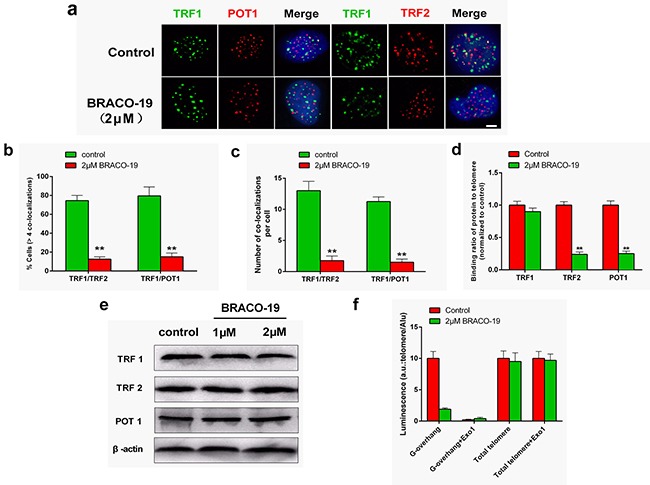
a. U87 cells treated with BRACO-19 (2 μM) for 72 hours were double stained with the indicated antibodies. Representative confocal images showing merged TRF1 (green) with TRF2 and POT1 (red) staining in untreated and treated cells. Scale bar equals 5 μm. b. Percentages of cells with more than four co-localizations per nucleus of TRF1/TRF2 and TRF1/POT1. Error bars indicated s.d. **P < 0.005. c. Average number of co-localizations per nucleus in U87 cells treated with BRACO-19 (2 μM). Error bars indicated ± s.d. **P < 0.005, two-tailed student's t-test. d. Binding of TRF1, TRF2 or POT1 was examined by ChIP assay and detected by qRT–PCR amplification of the telomeric region in U87 cells treated with BRACO-19 (2 μM). Data represented triplicate ChIP experiments, each with technical triplicates of qRT–PCR; **P < 0.01 as compared with controls. e. Expression of TRF1, TRF2 and POT1 in U87 cells treated with BRACO-19 (2 μM). β-actin was used as loading control. f. Hybridization protection assay (HPA) was performed on genomic DNA isolated from U87 cells treated with BRACO-19 (2 μM) to assess the length of G-overhang and total telomere length. ExoI nuclease digestion was used to assess integrity of the 3′-overhang. Luminescence intensity in arbitrary units (AU) was normalized against Alu probe. Error bars indicated ± s.d., **P < 0.01, two-tailed student's t-test.