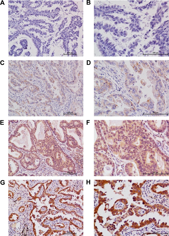
Figure 2. Immunohistochemical stainings of plakoglobin in the primary tumor of surgically resected lung adenocarcinoma.
A. A tumor tissue showed a negative staining of plakoglobin in all the tumor cells (200 ×). B. A tumor tissue showed a negative staining of plakoglobin in all the tumor cells (400 ×) (the IRS of this field: 0). C. the low staining of plakoglobin expression was detected in a tumor tissue (200 ×). D. The low staining of plakoglobin expression was detected in a tumor tissue, in which about 90% of tumor cells were observed (400 ×) (the score of this field: 4). E. the intermediate staining of plakoglobin expression was detected in a tumor tissue (200 ×). F. The intermediate staining of plakoglobin expression was detected in a tumor tissue, in which about 95% of tumor cells were observed (400 ×) (the IRS of this field: 8). G. the high staining of plakoglobin expression was detected in a tumor tissue (200 ×). H. The high staining of plakoglobin expression was detected in a tumor tissue, in which about 95% of tumor cells were observed (400 ×) (the IRS of this field: 12).

