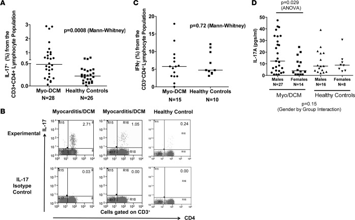
Figure 1. Th17 immunophenotype contributes to myocarditis and dilated cardiomyopathy (DCM).
(A) Peripheral blood mononuclear cells (PBMCs) from myocarditis/DCM subjects (n = 28) (baseline blood sample) or healthy controls (n = 26) were stained for CD4, CD3, and IL-17 and analyzed using a flow cytometer. Mann-Whitney, P = 0.0008. (B) Representative CD3+CD4+IL-17+ Th17 FACS diagrams are shown from myocarditis/DCM (n = 2) and a healthy control (n = 1). (C) Th1 (IFN-γ+) cells were not significantly high in myocarditis/DCM as a group in baseline blood samples. PBMCs from myocarditis/DCM subjects (n = 15) and healthy controls (n = 10) were stained for CD4, CD3, and IFN-γ and analyzed using FACS. Mann-Whitney, P = 0.72. (D) IL-17A in myocarditis/DCM males (n = 27) was significantly elevated at baseline compared to myocarditis/DCM females (n = 14) and trended toward a significant interaction by case/control status. Healthy controls: males (n = 16), females (n = 8). Three male and 4 female myocarditis/DCM samples had undetectable IL-17A and were placed on the graph halfway between zero and the lowest detectable value of 1.5 pg/ml. Two-way ANOVA, P = 0.029 (gender by group interaction, P = 0.15). FACS analysis was performed on fresh PBMCs, which were analyzed immediately upon receiving the blood sample. Only 1 sample per time point was analyzed by FACS and compared to isotype controls. Cytokine analysis was performed in triplicate to determine the serum cytokine concentration.

