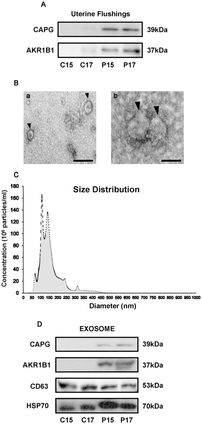
Fig 4. Identification of exosomal CAPG and AKR1B1 in days 15 and 17 pregnant UFs.

(A) The presence of CAPG and AKR1B1 in C15, C17, P15, or P17 UFs (10 μg each) was examined by western blot analysis (n = 3 each day). (B) Transmission electron microscopy analysis revealed the presence of approximately 150 nm vesicles in UFs, consistent with exosomes. Scale bar = 200 nm (a) or 100 nm (b). (C) Nanoparticle tracking analysis (n = 3, triplet analysis in each sample) of P17 UF revealed that a range of exosomal size is 50 to 150 nm. Gray area in (C) represents an average from three samples, whereas black area represents SEM. (D) Western blot analysis showed the presence of CD63 and HSP70 in exosomes isolated from C15, C17, P15, or P17 UFs, and the presence of CAPG and AKR1B1 in exosomes isolated from days 15 and 17 pregnant UFs (n = 3 each day). In (A), (B), and (D), a representative one is shown.
