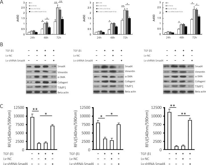
Fig 2. Effects of Smad4 knockdown on TGF-β1-induced proliferation and fibrotic markers in L6, RSC-364 and RC cells.
(A) Cell proliferation of the three cell lines and respective Smad4-silenced cell lines cultured in the presence or absence of TGF-β1 at the indicated time points. (B) Smad4, Vimentin, α-SMA, collagen I and Timp1 proteins expressed in L6, RSC-364 and RC cells and the respective Smad4-silenced cells induced by TGF-β1 were detected. (C) MMP activity was measured in the three cell lines and respective Smad4-silenced cell lines. Left, middle, right show the results from L6, RSC-364 and RC cells, respectively. The results are shown as the mean ± SD of at least 3 separate experiments. * P < 0.05 and ** P < 0.01 compared to the indicated group.

