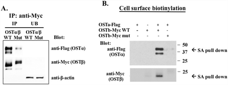
Fig 3. Co-Immunoprecipitation and surface biotinylation of the wild-type (WT) or LL/AA mutant (Mut) OSTβ-Myc with OSTα-Flag.
(A) The total protein extracted from transfected cells was immunoprecipitated by using anti-Myc proteinA/G agarose beads. The precipitates were separated by SDS-PAGE, and analyzed by Western blot using anti-Flag antibody to detect OSTα and anti-Myc antibody to detect OSTβ. (B) HEK293 cells were transiently transfected with WT or Mut OSTβ-Myc and OSTα-Flag. The OSTα and β proteins expressed on the cell surface were biotinylated and subjected to streptavidin agarose (SA) pull-down. The streptavidin agarose-bound proteins were separated by PAGE and blotted with anti-Flag or anti-Myc antibodies. The LL/AA OSTβ mutant prevents both subunits from going to the cell surface (lane #4).

