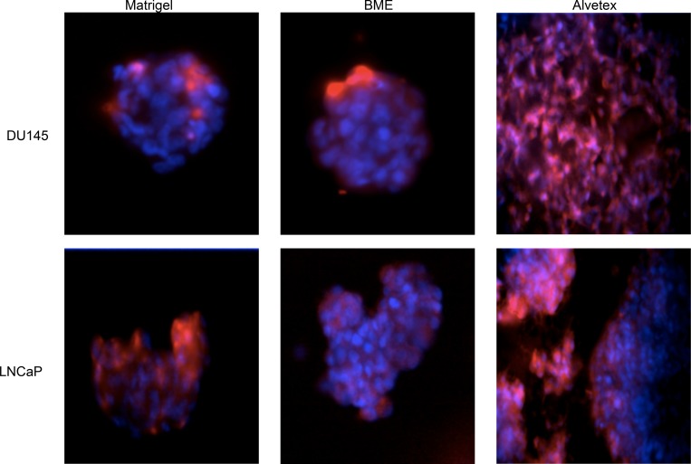
Fig 3. Fluorescent microscopic images of DU145 and LNCaP cells cultured in the 3D matrices: Matrigel, BME, and Alvetex.
Cells were seeded onto the various matrices and cultured for four days. Hoechst 33342 (blue) and CellTracker Red CMPTX dye (red) was used to stain nuclei and cytoplasm, respectively. Images obtained using BD Pathway Bioimager.

