Figure 3.
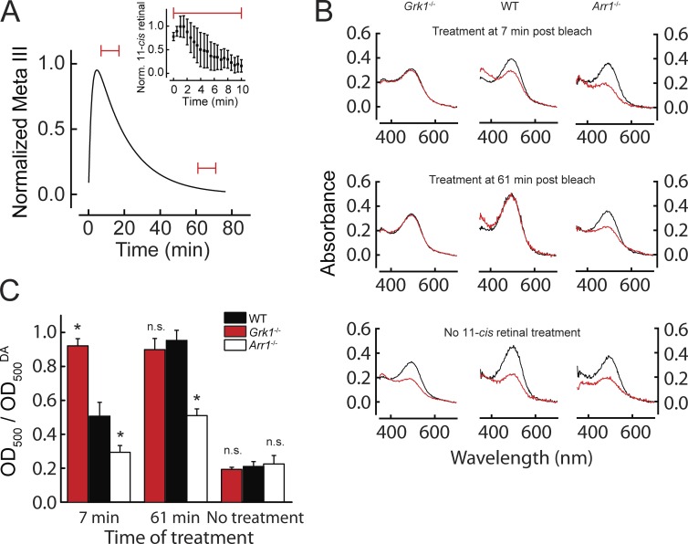
Pigment regeneration depends on Meta III decay in Grk1−/− and Arr1−/− mouse rods. (A) Typical experiment in which retinas were exposed to exogenous 11-cis retinal for 10 min (red bars) at either 7 min after bleaching (when Meta III reached its maximum) or 61 min after bleaching (Meta III decayed). The inset shows the change in concentration of 11-cis retinal in the recording chamber over the time of treatment normalized to its maximal concentration (∼10 µM). Data were reproduced from Fig. 4. (B) Spectra of Grk1−/−, WT, and Arr1−/− retinas measured 120 min after 80% pigment bleaching. The retinas were exposed to 10 µM exogenous 11-cis retinal for 10 min either at 7 min after bleaching when Meta III had reached its maximum (top) or 61 min after bleaching when there was no Meta III left in WT and Grk1−/− rods but a substantial amount left in Arr1−/− rods (middle). Bottom panels show untreated controls. Black traces represent prebleach spectra. Red traces show spectra measured after bleaching and/or treatment with 11-cis retinal. (C) Summary of the averaged data ± SEM from experiments as in B. The bars show the amount of pigment regenerated in WT (7 min, n = 4; 61 min, n = 4; no treatment, n = 7), Grk1−/− (7 min, n = 6; 61 min, n = 4; no treatment, n = 2), and Arr1−/− (7 min, n = 5; 61 min, n = 6; no treatment, n = 6) retinas when treated with 11-cis retinal 7 or 61 min after bleaching or not treated. Asterisks indicate a significant difference (P < 0.05) from WT; n.s., not significant (one-way ANOVA with Tukey’s test for means comparisons; F = 41; P = 1.3 × 10⁻¹⁵). Additionally, all models at both 7 and 61 min were significantly different from the untreated control, except Arr1−/− treated at 7 min. All recordings were made at 37°C.

