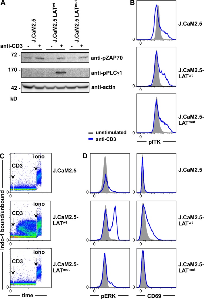
Figure 3.
TCR-induced signaling in LATmut T cells. (A–D) All experiments were performed in LAT-deficient J.CaM2.5 cells and J.CaM2.5 cells reconstituted with LATwt or LATmut. (A) Immunoblot of ZAP70 and PLCγ1 phosphorylation with or without stimulation with 5 µg/ml anti-CD3 for 3 min. (B) Phosphorylation of ITK (pITK) with or without stimulation with 5 µg/ml anti-CD3 for 2 min. (C) Ca2+ mobilization after anti-CD3 stimulation. (D) Phosphorylation of ERK (pERK) after 2-min anti-CD3 stimulation and up-regulation of CD69 after overnight stimulation. All results are representative of three to five independent experiments.

