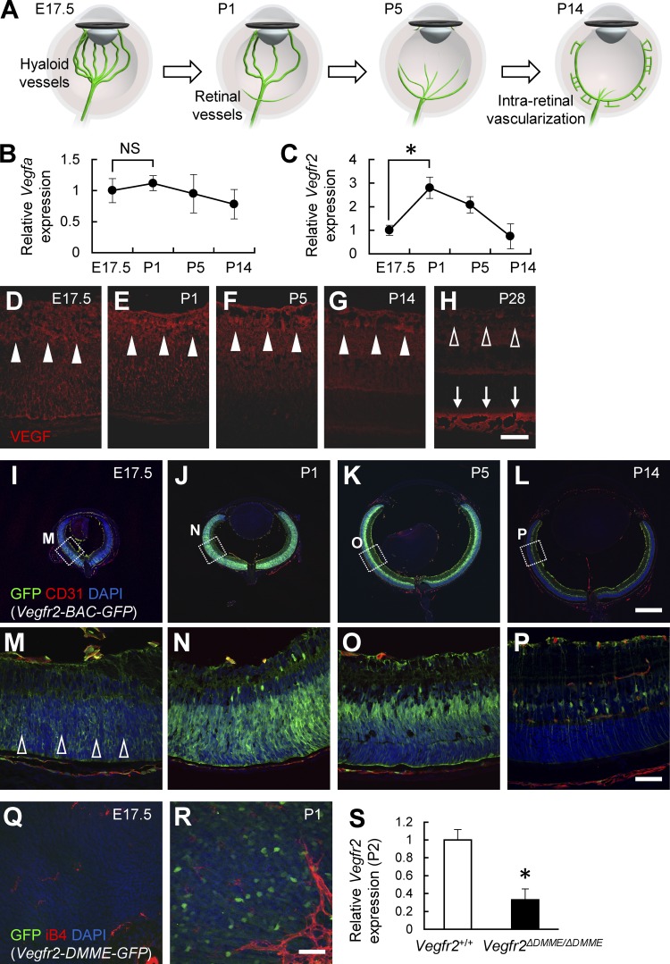
Figure 3.
VEGFR2 is markedly up-regulated via DMME in retinal neurons after birth. (A) Schematic diagram depicting the developmental transition of the ocular circulatory system. (B and C) Quantitative PCR analysis of retinas at P6 (n = 4). (D–P) Immunohistochemical analysis of retinal sections. VEGF was consistently detected in retinal neurons (closed arrowheads in D–G), although its expression decreased at P28 (open arrowheads in H). Abundant VEGF proteins were detected in the outer segments of P28 eyes (arrows in H). VEGFR2 expression in the embryonic day 17.5 (E17.5) retina is much weaker than that in the postnatal retinas (open arrowheads in M). (Q and R) Whole-mount retinas. (S) Quantitative PCR analysis of peripheral retinas at P2 (n = 4). Bars: (I–L) 500 µm; (D–H and M–R) 50 µm. *, P < 0.05; two-tailed Student’s t tests. Representative confocal images from three independent experiments (three mice per group) are shown. Data are represented as mean ± SD.

