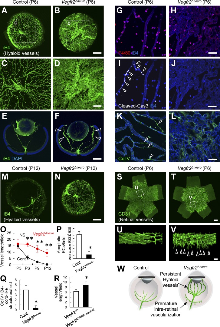
Figure 4.
Lack of neuronal VEGFR2 leads to persistent hyaloid vessels. (A–R) Whole-mount or section staining of hyaloid vessels at P6 or P12 and quantification (n = 4). The amount of hyaloid vessels is increased in Vegfr2Δneuro mice (arrowheads in F). The abundant endothelial apoptosis (arrowheads in I) and vessel regression (arrowheads in K) detected in control mice are greatly reduced in Vegfr2Δneuro mice. (S–V) Whole-mount staining of the retinal vasculature. Premature intraretinal vascularization (arrowheads in V) are seen in Vegfr2Δneuro mice. (W) Schematic diagram depicting vascular abnormalities in Vegfr2Δneuro mice. Bars: (A, B, E, F, M, N, S, and T) 500 µm; (C, D, U, and V) 200 µm; (G–L) 50 µm. *, P < 0.05; **, P < 0.01; two-tailed Student’s t tests. Representative confocal images from four independent experiments (four mice per group) are shown. Data are represented as mean ± SD. ECs, endothelial cells; ColIV, collagen type four; Cont, control.

